| Health Lecture |
 |
Medical quality concept build love human health
|
|
|
|
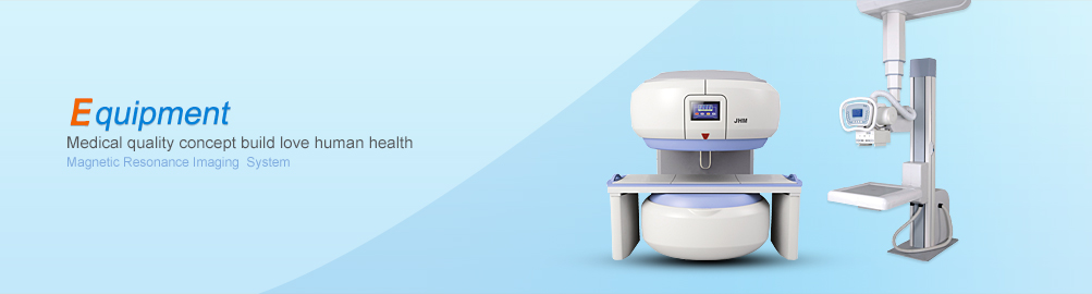
Magnetic resonance imaging (Nucler Magnetic Resonance Imaging referred MRI), is a diagnostic imaging technique eighties of last century was developed, because it completely out of the ionizing radiation on the human body damage, there are many parameters, the amount of information can be multi-faceted imaging, as well as a prominent feature of the soft tissue, such as high-resolution, then pay attention to all aspects of scholars from it come out, whether it is to improve the equipment, software updates and upgrades, or diagnosis of body parts and organs for research, developing very fast, now mature, is widely used in clinical diagnosis, for some diseases become an indispensable method of examination.
One, MRI is the use of the body proton nuclear magnetic resonance phenomenon occurs in the hydrogen-containing magnetic field, the MR signal collected, and then through the space constituting the image encoding technique, for doctors to do diagnosis. MR scanning equipment: magnets can be divided based on the formation of a permanent magnet type (lodestone constitution), and superconducting electromagnetic type three.
MRI equipment basic elements:
1. Magnet: In addition to the several sub-type, there are barrel closed type and open, the latter feasible intervention.
2. The gradient magnetic field: designed for spatial encoding, software function depending on its intensity and rate of change.
3. The RF coil: a plurality of types, transmit and receive RF pulse.
4. Acquisition System: Programs and imaging.
5. Computer: Requires large capacity, fast operation, full-featured, easy to operate.
Two, MRI features:
1.Grayscale Imaging: Like the X-ray, CT images are black and white, like the gray, but not the density, but the strength of the signal.
2. the flow void: liquid flow can not get a signal, no signal was in contrast with the surrounding signals, such as blood vessels, the flow of cerebrospinal fluid space.
3. can be multi-faceted, multi-dimensional imaging, two-dimensional, three-dimensional display of human anatomy and disease, not only to achieve localization diagnosis, diagnosis is also important reference value.
4. The amount of information, commonly used spin echo procedure, there are three basic image, namely as proton density, T1-weighted images, T2-weighted images, there are a variety of other imaging techniques, such as the use of blood flow void may constitute a flow imaging, angiography without contrast agent made, called "magnetic resonance angiography" MRA (MR Angio graphy), controlled by the body of water pipes made of the image called water imaging, such as bile duct imaging, renal pelvis and ureter imaging, spinal imaging and get rid of fat in order to observe the lesions with high signal interference called fat-suppressed image made imaging (STIR), there is also water suppression (FLAIR), and functional imaging studies, such as the human body functions. In conclusion, MRI can provide a lot of information and analysis for medical diagnosis.
5. since the nuclear magnetic resonance phenomenon, and directly reflects the state of the molecular structure of the surrounding environment of water molecules in the human body in the position of proton, which provides information on the biochemical and pathological conditions on the molecular level, allowing the human body edema, infection, inflammation changes in morphology and later formed on the degeneration prior to the early diagnosis, or super early diagnosis. This is the X-ray, CT, B ultrasonic imaging technology unparalleled.
6. a large soft tissue contrast, high resolution, determining inflammation, edema, tumors and other lesions is very clear, especially for surgery to determine the extent of surgery provides a very reliable basis.
7. there is no radiation damage to the human body, you can check multiple (multi-site, reviewed several times).
8. the majority of the cases do not need to use contrast agents, contrast agents currently used for a few cases the metal gadolinium chelates, called gadolinium diethylenetriamine pentaacetic acid di meglumine salt (abbreviated Gd-DTPA) is very safe, So far more than 20 years with no serious reactions reported death.
Three,MRI examination indications:
1. nervous system disease: cerebral infarction, brain tumors, inflammation, degeneration, congenital malformations, trauma, etc., for the application of the first body system, now has accumulated a wealth of experience, the lesion localization, diagnosis more accurate, timely, detect early lesions.
2. cardiovascular system: can be used to diagnose heart disease, cardiomyopathy, cardiac tumors, pericardial effusion, and mural thrombus, intimal flap peel like.
3. chest lesions: within mediastinal tumor, lymph nodes, and pleural disease, etc., can be displayed and relationships with large clumps of pulmonary vascular trachea.
4. abdominal organs: liver cancer, liver hemangioma liver cyst diagnosis and differential diagnosis of abdominal tumor diagnosis and differential diagnosis, especially retroperitoneal lesions.
5. pelvic organs; uterine fibroids, other tumors, ovarian tumors, qualitative positioning within the pelvic mass, rectum, prostate and bladder tumor and so on.
6. Bone and joint: bone infections, tumors, trauma diagnosis and extent of disease, especially for some minor changes, such as a bone bruise and so have a greater value, intra-articular cartilage, ligaments, menisci, synovium, synovial capsule, etc. lesions and bone marrow lesions have a high diagnostic value.
7. The body of soft tissue lesions: whether from nerves, blood vessels, lymphatic vessels, muscle, connective tissue tumors, infections, degenerative diseases, etc., can be made more accurate positioning, qualitative diagnosis.
Fourth, check Notes:
1. Install artificial pacemakers and neurostimulators who were prohibited for tests.
2. intracranial silver clip and intraocular metallic foreign bodies were prohibited for tests.
3. ECG can not enter the MRI examination room. Have done artery disease surgery, heart surgery and have done with artificial heart valves were prohibited for tests.
4. All kinds of critically ill patients: such as trauma or coma after an accident, irritability, heart disorders, respiratory insufficiency, continuous bleeding and stool incontinence and more.
5. Check the parts of a metal object (such as a fixed needle nails, etc.) can not be checked.
6. Pregnant women do careful examination, if possible pregnancy, please inform the examining physician.
7. Keep medical records, X-ray, CT films, films and other materials that came with previous MRI bring MRI room for reference. |
|