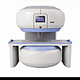
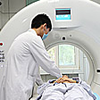
MRI images and CT images are very similar, both are "digital image", and gradation display different cross-sectional images of different anatomical and pathological structures. Like with CT, magnetic resonance imaging is also suitable for almost all body systems of different diseases, such as examination of tumor, inflammation, trauma, degenerative disease, and a variety of congenital diseases like.
Magnetic resonance imaging artifacts boneless, free for direct multi-direction (transverse, coronal, sagittal, or any angle) cut layer of brain, spine and spinal cord, such as anatomy and lesions showed more preferable range in CT, Magnetic resonance imaging by means of its "flow void", can not angiography agent, showing the vascular structure, it displays vessels (except the tiny blood vessels), as well as mutual authentication between the tumor, lymph nodes and vascular structures in the "no damage" to aspects, unique. Magnetic resonance imaging of soft tissue CT several times higher than the resolving power, it can be sensitive to the detected change in the water content of tissue components, so often more effective than CT and found early lesions. In recent years, magnetic resonance flow imaging technology, the in vivo measurement of blood flow and blood flow velocity has become possible; the use of ECG-gated, so that MRI can clearly and comprehensively show the heart, myocardium, pericardium and other small structures inside the heart,For non-destructive examination and diagnosis of various acquired and congenital heart disease (including coronary heart disease, etc.), and to check heart function, providing a reliable method. With a variety of fast scan sequence and three-dimensional scanning technology research and sampling successfully used in clinical, magnetic resonance angiography and cinematography new technology has entered clinical and improving. Recently, magnetic resonance imaging and to achieve a combination of the local spectrum Science (i.e. the combination of MRI and MRS), as well as other nuclei such as fluorine, sodium, phosphorus magnetic resonance imaging other than hydrogen protons, these achievements will be more effective in improving MRI diagnostic specificity, but also broaden its clinical use.
Magnetic Resonance Imaging of the main problems is that the time it takes longer to scan, so check for some patients often do not match the sense of difficulty, sports sexual organs, such as the gastrointestinal tract due to the lack of suitable contrast agent is often difficult to read ; for the lungs, due to breathing exercises and alveolar proton density is low and other reasons, the image is not satisfied. Magnetic resonance imaging calcifications and bone lesions show, not as accurate and sensitive CT. Magnetic Resonance Imaging spatial resolution room, also needs to be further improved.
1. The brain and spinal cord MRI diagnosis of brain tumors, encephalitis lesions, white matter lesions, cerebral infarction, cerebral congenital abnormalities is more sensitive than CT, can detect early lesions, positioning is more accurate. On the base of the skull and the brain stem lesions because no artifacts can be displayed more clearly. MRI contrast agents can not show cerebrovascular, discover whether the aneurysm and arteriovenous malformations. MRI also directly show some cranial nerves, can be found on these nerves occurred early lesions. MRI can directly show the whole picture of the spinal cord, thus the spinal tumor or spinal tumors, spinal cord white matter lesions, syringomyelia, spinal cord injury and other important diagnostic value. For disc disease, MRI can show its variability, prominent or bulging. Display spinal stenosis better. For the neck, spine, CT often show satisfied, while MRI showed clearly. In addition, MRI for vertebral metastases are also very sensitive display.
2. Department of Otolaryngology Head and Neck MRI eye Ministry neoplastic lesions show well, such as nasopharyngeal carcinoma of the skull base, cranial nerve violations, MRI display clearer and more accurate than CT. MRI angiography of the neck can be done to show vascular abnormalities. Of the mass of the neck, MRI can also display its range and features to help qualitatively.
3. chest MRI can show direct myocardial and left ventricular cavity (intentions gated), can understand the situation of myocardial damage and cardiac function was measured. Mediastinal large vessels of the situation can be clearly indicated. Mediastinal tumor localization and characterization is also very helpful. Display cases also pulmonary edema, pulmonary embolism, lung cancer. Distinguishing the nature of pleural effusion, or lymph vessel section difference.
4. Abdominal MRI in the diagnosis of liver, kidney, pancreas, spleen, adrenal glands and other organs of the disease can be substantial and provides very valuable information that can help confirm the diagnosis. Small lesions are easier to display, which can detect early lesions. MR pancreatic cholangiography (MRCP) to display the biliary and pancreatic ducts, and can replace ERCP. MR urography (MRU) to display the expansion of the ureter and renal pelvis, renal function is poor, the patient developed IVU not particularly applicable.
5. Pelvic MRI can show the uterus, ovaries, bladder, prostate, seminal vesicles and other organs of the disease. Can directly see the endometrium, myometrium, the early diagnosis of uterine cancer lesions of great help. Positioning diagnosis of ovarian, bladder, prostate disease, etc. is also a great value.
6. MRI of retroperitoneal tumors and peritoneal relations with the surrounding organs after displaying great value. Also show the abdominal aorta or other large vessel disease, such as abdominal aortic aneurysm, cloth - check syndrome, renal artery stenosis.
7. musculoskeletal MRI for intra-articular cartilage discs, tendon, ligament damage, display rate than CT. Because of the more sensitive to changes in the bone marrow, early detection of bone metastases, osteomyelitis, aseptic necrosis, leukemia bone marrow infiltration. Bone and soft tissue tumors block display clearly. Soft tissue injury also have some diagnostic value.
|